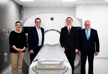


Some medical issues are very clear, a heart attack or a tumour for instance. But many medical issues and conditions have doctors looking for tears and lesions sometimes less than a millimeter in size — the health equivalent of finding a needle in a haystack.
The hunt for these critical issues can result in multiple invasive procedures, which are stressful for both the patient and the healthcare system.
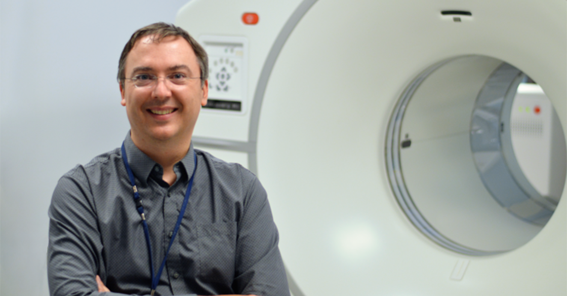
A photon counting CT (PCCT) scanner is the next generation of this essential scanning technology, which Dr. James Clarke, head of the department of diagnostic radiology at the QEII Health Sciences Center, says is rocking the diagnostic imaging world with new and efficient ways to address the issues patients face.
In this Q&A, Dr. Clarke tells us how the drive to bring a PCCT scanner to the QEII represents a transformative leap forward in diagnostic imaging in Atlantic Canada — redefining what clinicians can see and how quickly they can act.
______________
Q: What exactly is a photon counting CT scanner?
A: A photon counting CT is a new CT technology, emerging over the last few years where you can actually detect each x-ray photon as it arrives at the detector. Traditional CT scanners work with x-ray tubes that produce the x-rays, which pass through the patient. The tubes produce x-rays of many different energies – called a spectrum. A bunch of those x-rays get absorbed by the patient, and some of them reach the detector on the far side, allowing us to generate our pictures. In a traditional CT, we don’t know much about the x-rays that reach the detector. A photon counting CT is different because you can determine the characteristics of the x-
For a lot of studies, we administer intravenous contrast (some people call this X-ray dye) and we watch to see how that contrast moves through the patient's blood stream and organs. It shows up at different times in different places and that gives us information about things that are going on inside the patient. With photon counting CT, we can see the contrast better which allows us to use lower doses of both contrast and x-ray radiation.
Q: You mentioned the PCCT technology is new, but what impact has it had on the diagnostic imaging world?
A: It’s really exciting and medical professionals are still figuring out all of the applications for it and more applications are coming out all the time.
The best conventional CT scanners out there have a resolution that's about 0.6mm, which is already really small. But some of the structures that we're looking at in the body are also really small. As a cardiac radiologist, the coronary arteries we need to see that feed your heart muscle be as small as 1.5-2mm in size. So if you need to figure out if an artery is narrowed that can be really difficult to sort out.
Conventional CT also has problems with high density structures like metal. If you needed a stent to open up a blocked blood vessel, we couldn’t really see inside those tiny metal structures on a conventional CT. We would see this blurry thing that's high density but we couldn’t see inside it to see if it was blocked again.
The difference in the technology on the photon counting CT is that the detector directly absorbs the x-ray and turns it into an electrical signal within the detector, which leads to a much higher resolution. So instead of 0.6mm being your resolution for a traditional CT scanner, you can get down to 0.2mm resolution — three times higher spatial resolution. When you make it a 3D image, this means your effective resolution is 27 times better than a traditional CT scanner. It is amazing!
Q: Is this the next generation of CT scanners?
A: All CT scanner vendors are basically saying that within five to 10 years, almost every CT scanner that gets installed on the planet is going to be photon counting. We are at the leading edge of the wave.
Q: For doctors, gaining access to a PCCT is really arming you and the rest of the diagnostic imaging team to see all the potential problems and gain that better image. What about for the patient?
A: For the patient, there are new benefits because we have better scan resolution. We can start to solve problems that we weren't able to solve in the past.
One of the things that came up relatively early when people started working with PCCT scanners is the opportunity to address a condition called a cerebrospinal fluid leak — a condition where the fluid that cushions your brain and spinal cord escapes through a small tear. This can lead to all sorts of health problems, and they can be very difficult to detect because they're typically very small tears. There are invasive procedures to try and find these leaks but with the PCCT scanner, it’s “just” a scan.
This technology is also revolutionary for cardiac patients – being able to look inside those coronary stents and see if there's narrowing or blockages, potentially saving them from invasive procedures like cardiac catheterization. It also reduces the radiation exposure a patient may receive.
Q: Is there an opportunity for cancer patients to benefit from a PCCT scan?
A: Yes, specifically when it comes to characterizing lesions and looking at response to treatment, being able to more accurately measure sizes. The cancer side of PCCT development is happening a little later than other areas. There's a lot of work and research being done to look at how the spectral information that we're getting from the photon counting CT can potentially tell you how lesions are responding to treatment and then guiding therapy from that. There's active research going on to see how this technology can help improve cancer therapeutics and management.
Q: Overall, why do we need a PCCT at the QEII?
A: We need it because we're the centre of excellence for cardiac imaging — this is where we do our interventional cardiology diagnostics. The QEII is also where our centre for neurosurgery is. It’s our centre for innovation in diagnostic imaging. It’s our major trauma centre and the site where we also do research and education.
Having access to the latest technology and the technology of the future will allow us to train our technologists, residents, and all of our learners on what they're going to be working with in the years to come. State-of-the-art technology will allow us to do and be involved in exciting research projects and advances clinical care for patients at the QEII and across Nova Scotia.
So, this PCCT will be a workhorse! It's a very good CT, but it's also got all these extra bells and whistles and new things that it can do.
Q: What would you say to donors who are thinking about investing in this technology?
A: This is the technology of the future. This will be helpful for patients, our medical teams, and the healthcare system. It will allow us to be at the cutting edge of medical imaging in Canada and around the world. Having a PCCT scanner will bring us, at the QEII, to the forefront of CT imaging worldwide.
Making a gift to the QEII Foundation this holiday season can help bring the cutting-edge photon counting CT scanner to the QEII, Atlantic Canada’s most specialized hospital. You can make your donation today at QE2Foundation.ca/Diagnostic-Imaging