

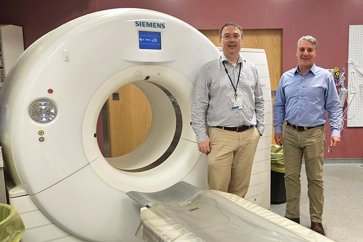
Dr. James Clarke (left), chief, Department of Diagnostic Imaging, Central Zone, and Dr. Jens Heidenreich (right), head of neuroradiology and head and neck sections at the QEII’s Department of Diagnostic Imaging, stand in front of the QEII’s old FLASH scanner, which will be replaced by the new photon counting CT. With enhanced images, the new scanner will provide better accuracy for the doctor diagnosing health conditions. CONTRIBUTED
QEII Foundation raising funds for state-of-the-art PCCT at the QEII
The QEII Foundation is raising funds to bring a state-of-the-art photon counting computed tomography (PCCT) scanner to the QEII Health Sciences Centre as part of its $100-million We Are campaign.
Like current CT scanners, the new technology takes X-rays from many directions in a circle around the patient, creating a 3D picture of what’s going on inside them but with greatly increased resolution. It will be especially important for heart and cancer care patients.
The enhanced images will provide better accuracy for the doctor diagnosing health conditions, which can lead to reduced wait times for other imaging scans by helping answer questions with a single study instead of multiple studies. The new scanner will also decrease radiation exposure for patients. There are currently only three Canadian hospitals using the advanced PCCT scanners, all in Vancouver and Toronto.
Dr. James Clarke is the chief of the Department of Diagnostic Imaging, Central Zone, Nova Scotia Health.
“It's an entirely new technology,” he says. It will overcome some of the hard physical limitations of the older CT scanners. Those devices can only see at 0.6 millimetres when studying a patient’s blood vessels. If you are trying to figure out what is going on in a two-millimetre blood vessel, it can make it hard for doctors to decide on a next step.
Dr. Clarke says the photon-counting scanner goes down to 0.2 millimetres, giving them three times the spatial resolution.
“It allows you to more definitively and more clearly measure those vessels, measure the narrowings in those vessels, and see whether or not there's something that someone needs to be worried about,” he explains.
The new machine also gives them information about the energy of the X-rays that make it through the patient. That helps overcome issues with older CTs that have problems dealing with metal in the patient, which absorbs those X-rays.
“The photon-counting CT fixes a whole bunch of those problems. We can see inside the metal stents that a cardiologist would use to fix someone's artery.”
It also lets them look at blood vessels in the brain for patients who have aneurisms. “That 0.2 millimetres lets us better characterize and better identify aneurisms in the brain, which can be catastrophic if they rupture.”
He hopes it will allow them to see things clearly the first time and reduce the need for more invasive followup procedures or additional testing.
Dr. Clarke says patients won’t notice much difference and will still lie on a bed as the “big donut” machine whirls around them. It also reduces radiation exposure to the patient, which can add up if you need a lot of scans.
“A big thanks to QEII Foundation donors for supporting imaging technology and keeping the hospital at the forefront. This is going to be a gamechanger for patients.”
Dr. Jens Heidenreich, who leads the neuroradiology & head and neck sections at the QEII’s Department of Diagnostic Imaging, says it will be a huge win for patients across Atlantic Canada. He remembers the old scanners twenty-five years ago that took twenty minutes to study one person. This generation of machines needs only seconds. He says it’s a big step forward.
“We have, over the years, moved further and further along from purely anatomical imaging to more functional imaging where we try to understand the underlying biology better,” he says.
He says the new machine will help doctors see if chemotherapy is working on the tumour cells on a molecular level.
“We do imaging of the central nervous system. We image the brain, the head and the spinal cord,” he says. “We look at these tiny little structures that conduct the hearing and we look at how the hearing gets processed in the inner ear and in the middle ear and all the imaging in the soft tissues of the neck.”
Dr. Heidenreich says the new machine’s higher detail resolution could, for example, better detect a blood clot in the brain that could be removed to save a life.
“Even we as highly trained radiologists, we can't look beyond certain levels, but obviously very fast computers can detect subtle differences that we can't detect,” he says.
The machine is expected to be installed by spring 2027.
To learn more or to help fund the PCCT, click here.